
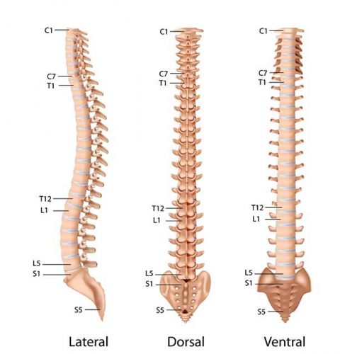

Scoliosis or lateral deviation of the lumbar spine
The curvature or deviation of the vertebral column is interpreted as scoliosis What is scoliosis? This complication is a lateral deviation of the vertebral

What is a brain tumor and what are its complications?
A brain tumor is an abnormal growth of brain cells or surrounding tissues. What is a brain tumor? Brain Tumor Growth and expansion
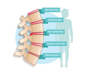
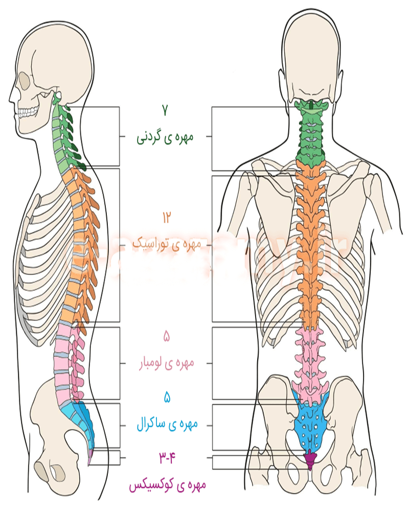
What diseases can the spine be exposed to?
Diseases and disorders of the spine The spine is an important part of the human skeletal structure and is composed of vertebrae, discs, muscles,
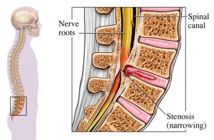
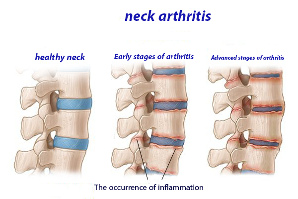
The latest research in the field of spinal canal stenosis, causes, symptoms and treatment methods
What is spinal stenosis? Spinal stenosis is a medical condition that results in blockage or narrowing of the spinal canal. This spinal canal is